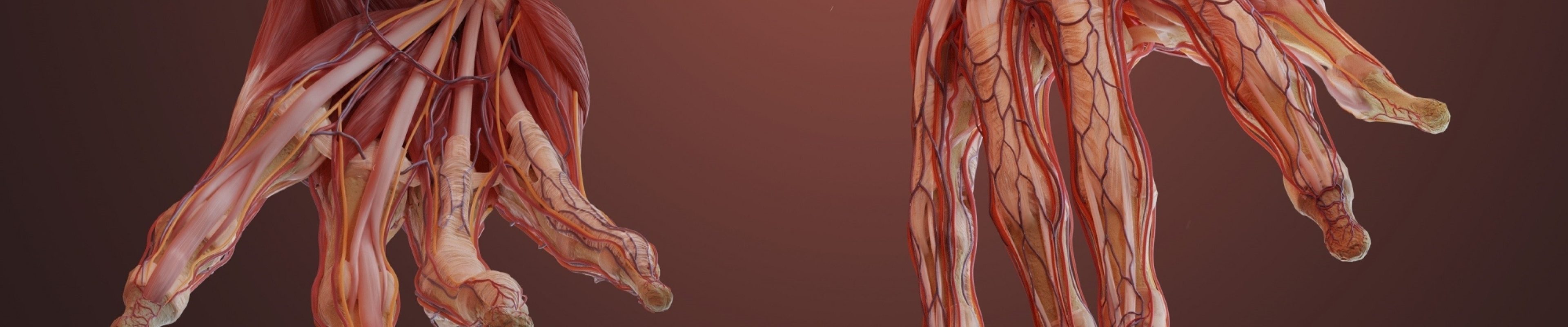
Illustration of a superficial dissection of the forearm by Matt Briggs
Body Parts: the art and application of medical illustration
Open Tuesday - Saturday, 10am - 5pm
20th September–9th November 2024
The tradition of making images of the human body for medical education goes back centuries. The development of anatomy atlases, so called as they 'mapped' the body began in the 1400s. Manuals for surgery and medical treatments followed.
Today original illustrations might be drawn in pencil, ink or paints, or created in digital packages - all are made to communicate information about human anatomy, medical conditions and surgical procedures. They can be widely seen in healthcare information leaflets, public health posters and campaign materials, with further uses in forensic photofit portraits, visualising aging and plastic surgery applications. Diagrams for exercise and yoga books; models used to train healthcare students and clinicians and even museum exhibits - all help professionals and the public better understand health and communicate medical treatments.
Please note this exhibition may be closed for school sessions on Tuesdays and Thursdays between 10:00am - 12:15pm, and 1:45pm - 3:45pm. The Hunterian Museum will remain open.
Find out more about the Medical Artists' Association of Great Britain
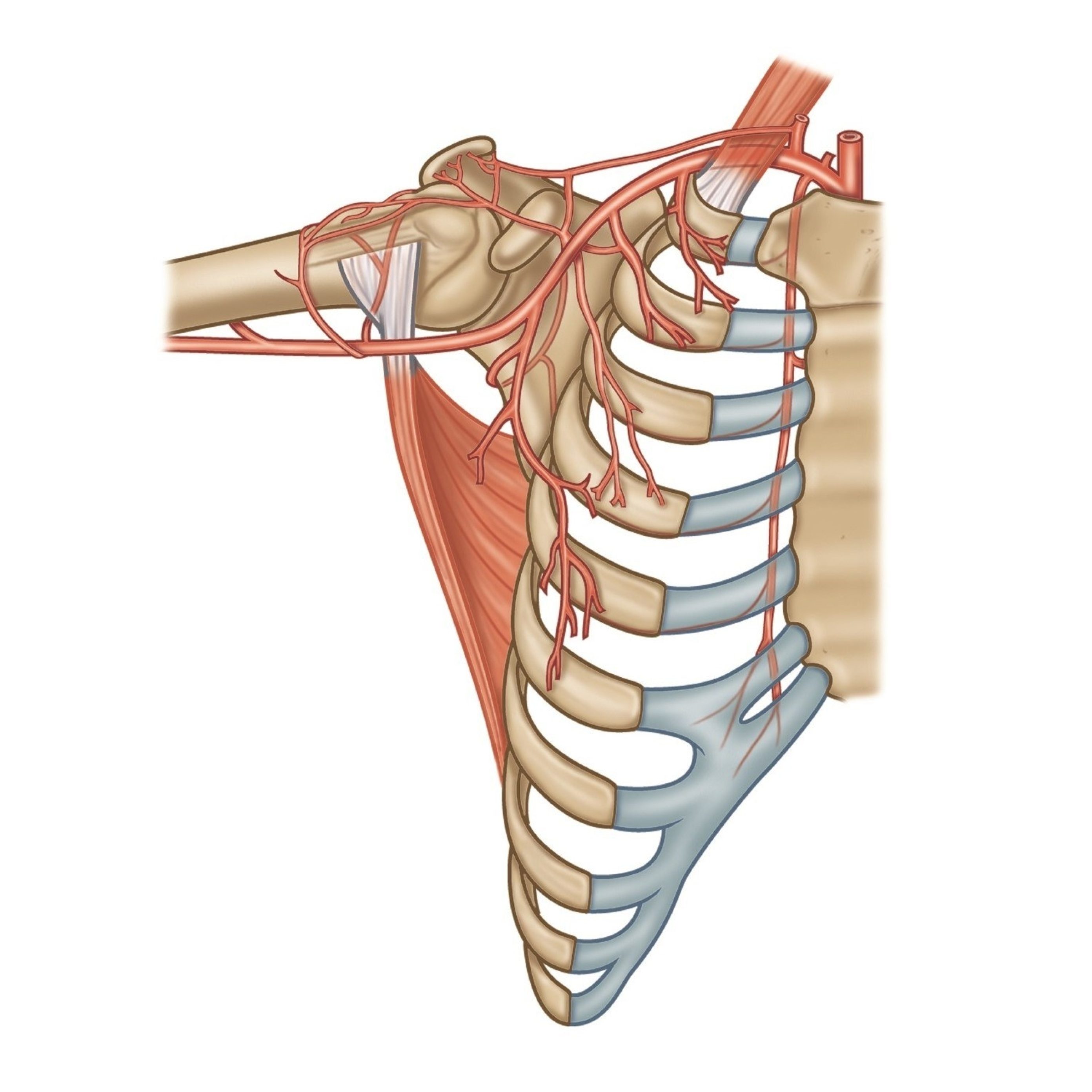
Arterial supply to the latissimus dorsi muscle by Amanda Williams

Coronavirus by Maurizio De Angelis
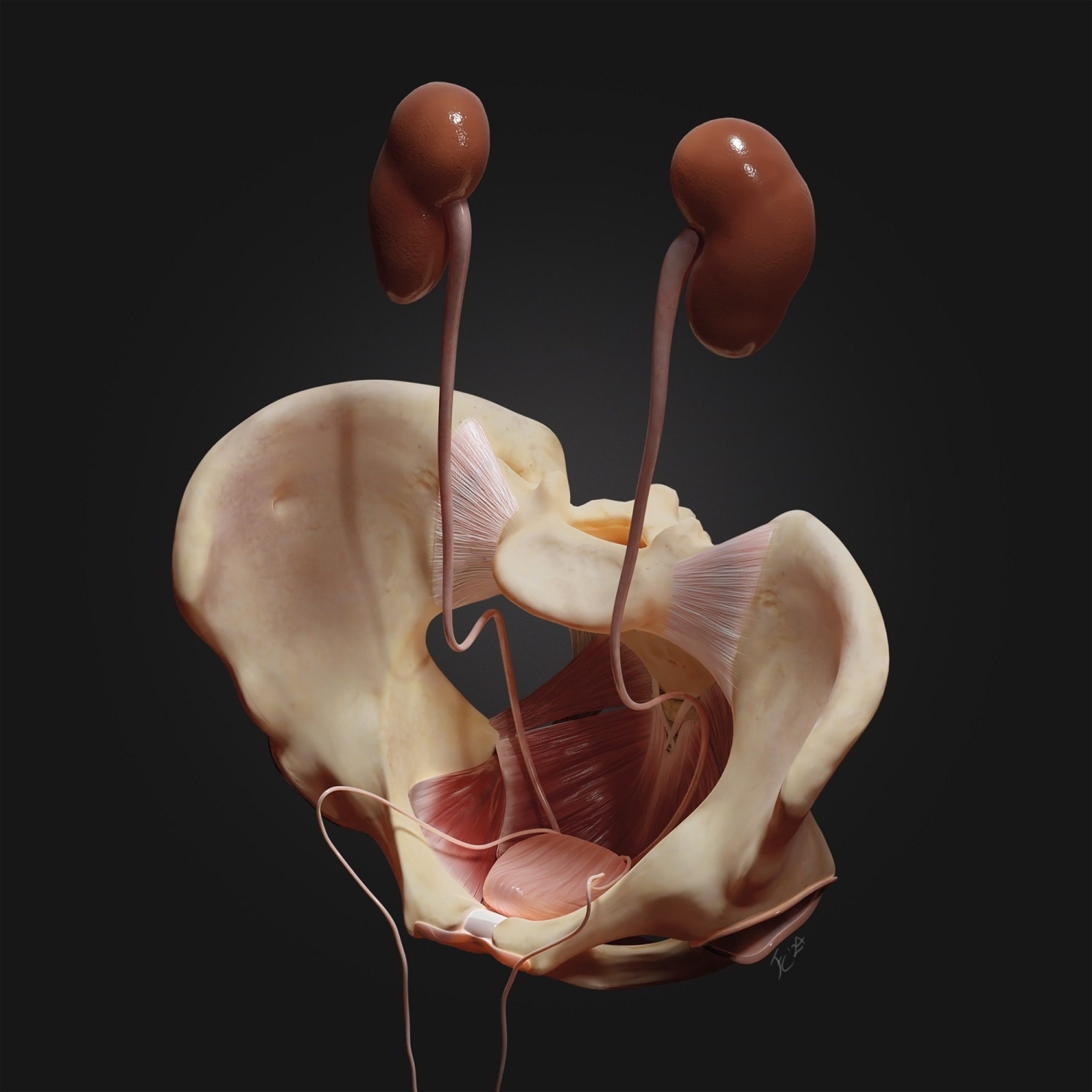
Urinary system by Francesca Corra

Growing uterus stages by Joanna Butler