New Hunterian Museum displays, Room 1
Hunterian Museum displays
The Hunterian Museum is named after the 18th century surgeon and anatomist, John Hunter (1728-1793). It includes the display of over 2,000 anatomical preparations from Hunter’s original collection, alongside instruments, equipment, models, paintings and archive material, which trace the history of surgery from ancient times to the latest robot-assisted operations. The Museum includes England’s largest public display of human anatomy.
Layout of the Hunterian Museum
Room 1
Hunterian Museum - an introduction
A journey through the museum begins with an introduction to the Royal College of Surgeons of England's museum collections. The Hunterian Museum, with John Hunter’s collection of 14,000 anatomical specimens at its heart, first opened in 1813. Thousands of anatomy and natural history preparations, surgical instruments, models, paintings, drawings and other objects have been added since. This space displays highlights from these collections.

Private Thomas Walker, by Thomas William Wood, 1856
Surgery and Anatomy - from ancient times to the 1700s
Surgery has been practiced throughout the world for thousands of years. This area charts how surgical treatments and anatomical knowledge have developed over centuries.
This space displays the Evelyn Tables, made from real human tissue. Anatomists carefully dissected blood vessels and nerves from bodies and pasted the tissue onto wooden boards. Dating from the 1640s, they are the oldest surviving anatomical preparations of their kind.

The Evelyn Tables
Room 2
John Hunter - A Curious Mind
John Hunter - the 18th century surgeon and anatomist who gives the museum its name - is introduced in this room. Born in Scotland in 1728, he received little formal education as a child; Hunter’s learning and inspiration came instead from observing and questioning the natural world around him.
When I was a boy, I wanted to know all about the clouds and the grasses, and why the leaves changed colour in the autumn; I watched the ants, bees, birds, tadpoles, and caddisworms; I pestered people with questions about what nobody knew or cared anything about.

Bumblebees, prepared by John Hunter, 1760-1793

Twig showing small flowers, prepared by John Hunter, 1760-1793
Room 3
John Hunter - The Making of a Surgeon
How did John Hunter become a surgeon? This room explores Hunter's early career, working at his brother William's anatomy school before enlisting as an army surgeon, his partnership with the dentist James Spence, and appointment as surgeon to St George’s Hospital.

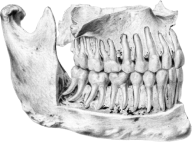
Human teeth, prepared by John Hunter, 1760-1793
Room 4
The Long Gallery - John Hunter's Collection
The majority of John Hunter's collection - his life's work - is displayed in this gallery. Made from human, animal and plant tissue, they show how he understood the natural world.
Room 5
John Hunter - Earl's Court
By 1765, John Hunter was earning enough to buy a small country estate at Earl’s Court, a village west of London. This room looks at some of the work he carried out there - observing nature, setting up experiments, dissecting animals and preparing specimens.

Love-in-a-mist (Nigella) seed pod, prepared by John Hunter, 1760-1793
Room 6
John Hunter - Leicester Square
In 1783 John and his family moved to a new home in the center of London. The property combined two houses: 28 Leicester Square and 13 Castle Street. The front was a fashionable town house, the back Hunter’s work rooms and anatomy school. The displays in this room explore the differences between these spaces, and how Hunter acquired specimens.

Boar’s epididymis (tube which stores and transports sperm) injected with mercury and framed for display, prepared by John Hunter, 1760–93
Room 7
John Hunter - St Georges Hospital
John Hunter’s patients ranged from the poor to the wealthy. He served as a surgeon at London’s St George’s Hospital for 25 years, and had a private practice treating people in their own homes. This space tells the stories of a some of his patients - their lives, professions, illnesses, treatments and in some cases their deaths.
John Hunter died in 1793. The displays in this room goes on to look at the students he influenced, and the development of the Royal College of Surgeons of England collection after his death.

John Burley, aged 37, who had a salivary adenoma - a benign tumour - growing in his jaw

John Burley after Hunter successfully operated to remove the tumour in 1785
Room 8
New Frontiers
The practice of surgery was transformed in the 1800s by three major breakthroughs which are explored in this gallery: the relief of pain, the introduction of germ-free surgical environments and the identification of disease at a cellular level. By 1900, surgeons were operating on every part of the human body.

Ivor Back FRCS (1879–1951) by William Orpen, 1926
Room 9
Modern Surgery
Dynamic and innovative, modern surgery continues to respond to the challenges of a complex world. This room looks at scientific advances from 1914 to 2023, and how they have transformed the way we understand and operate on the human body. The displays also explore what key developments and challenges the future may bring.

Akutsu artificial heart, 1981
Room 10
Transforming Lives
The final room looks at personal experiences of surgery, from both the patients’ and the surgeons’ perspectives. It tells of life saving and life enhancing surgery, and the forging of unique personal and professional bonds.
The Operation Room, 2022